Historical concepts of the athlete’s heart. Thyroid hormone signalling is altered in response to physical training in patients with end-stage heart failure and mechanical assist devices: potential physiological consequences? Interact Cardiovasc Thorac Surg. 2003 108:554–9.Īdamopoulos S, Gouziouta A, Mantzouratou P, Laoutaris ID, Dritsas A, Cokkinos DV, et al. Antiremodeling effect of long-term exercise training in patients with stable chronic heart failure: results of the Exercise in Left Ventricular Dysfunction and Chronic Heart Failure (ELVD-CHF) Trial. Giannuzzi P, Temporelli PL, Corrà U, Tavazzi L, ELVD-CHF Study Group. Low-intensity exercise training delays heart failure and improves survival in female hypertensive heart failure rats. 2002 54:162–74.Ĭhicco AJ, McCune SA, Emter CA, Sparagna GC, Rees ML, Bolden DA, et al. Aerobic exercise reduces cardiomyocyte hypertrophy and increases contractility, Ca2+ sensitivity and SERCA-2 in rat after myocardial infarction. Wisløff U, Loennechen JP, Currie S, Smith GL, Ellingsen Ø. Src and multiple MAP kinase activation in cardiac hypertrophy and congestive heart failure under chronic pressure-overload: comparison with acute mechanical stretch. Takeishi Y, Huang Q, Abe J, Glassman M, Che W, Lee JD, et al. QT dispersion and T-loop morphology in late pregnancy and after delivery. Lechmanová M, Kittnar O, Mlcek M, Slavícek J, Dohnalová A, Havránek S, et al. Molecular and functional signature of heart hypertrophy during pregnancy. 2002 97:73–8.Įghbali M, Deva R, Alioua A, Minosyan TY, Ruan H, Wang Y, et al. Left ventricular hypertrophy and diastolic dysfunction in healthy pregnant women. Schannwell CM, Zimmermann T, Schneppenheim M, Plehn G, Marx R, Strauer BE. Postprandial angina pectoris: clinical and angiographic correlations. Regional myocardial blood flow redistribution as a cause of postprandial angina pectoris. Ventilatory and cardiovascular responses of a python (Python molurus) to exercise and digestion. Physiology: postprandial cardiac hypertrophy in pythons. 2007 49:962–70.Īndersen JB, Rourke BC, Caiozzo VJ, Bennett AF, Hicks JW. The fuzzy logic of physiological cardiac hypertrophy. On the mechanism of compensatory hyperfunction and insufficiency of the heart. Wall stress and patterns of hypertrophy in the human left ventricle. Cardiac hypertrophy: the good, the bad, and the ugly. Phenotyping hypertrophy: eschew obfuscation.

Development and proliferative capacity of cardiac muscle cells. The possible therapeutic approaches are discussed. The increase in mass affects not only cardiomyocytes but cardiac fibroblasts, extracellular matrix, and endothelium and vascular smooth muscle cells. Various pathological genes appear that mediate this type of HYP, which to a large extent does not regress after removal of the noxious stimulus. Pathological or maladaptive HYP is more commonly seen in hypertension, valvular heart disease, and myocardial heart disease HYP can be marked, myocardial dysfunction occurs, and there is a return to the fetal phenotype, cell death, and fibrosis.


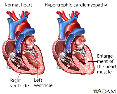
A return to the fetal phenotype or increase of fibrosis does not occur. HYP can be physiological and adaptive, commonly seen in pregnancy and exercise myocardial mass increase is moderate, cardiac contractility is normal or supernormal, and HYP regresses after child birth or detraining. In the clinical aspect, the left ventricle is the one more commonly affected and studied. Cardiac hypertrophy (HYP) is an increase of cardiac mass.


 0 kommentar(er)
